

The way stem cells distinguish themselves from other cells is one of the most fascinating processes I have studied in medical science. These master cells stand out as the body’s only cells that can turn into more than 200 different specialized cell types. They are quite remarkable — unlike regular cells, which have limited lifespans, stem cells keep renewing themselves and producing exact copies.
Stem cells are the building blocks of the human body, which uses them as its natural repair system. Hematopoietic stem cells maintain blood and immune cells, while mesenchymal stem cells support bone, cartilage, muscle, and fat. What makes these cells especially remarkable is their ability to differentiate into many different cell types — from blood cells to nerve cells and heart muscle cells. This makes them invaluable for medical research and treatment.
Stem cell therapy shows promise for treating a wide range of diseases. This is especially important because about 60% of American adults live with a chronic disease, highlighting a genuine need for new treatments. The list of conditions keeps growing — from arthritis and multiple sclerosis to various cancers and heart disease. These cells offer hope when conventional treatments do not work.
This article explores the science of cellular reprogramming and how scientists are using stem cells to transform medicine. We will examine what drives cell development, the most effective methods for guiding cell growth, and the promising clinical applications that are improving patient care.
Molecular Basis of Stem Cell Differentiation
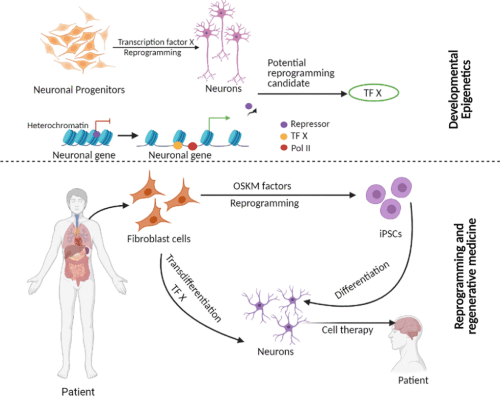
Image Source: Clinical Epigenetics – BioMed Central
The molecular complexity of stem cell differentiation depends on intricate genetic and epigenetic mechanisms that transform unspecialized cells into functional, tissue-specific cells. Scientists now regard understanding these mechanisms as essential to advancing regenerative medicine approaches.
Pluripotency vs Multipotency: Key Differences
A stem cell’s differentiation capacity defines its cellular potency. Pluripotent stem cells (PSCs) can differentiate into cells from all three germ layers—ectoderm, mesoderm, and endoderm. These cells cannot form extraembryonic tissues such as the placenta or umbilical cord. Multipotent stem cells differentiate only into closely related cell types within a specific lineage. Blood-forming stem cells (HSCs) produce exclusively blood cell types, while mesenchymal stem cells (MSCs) develop into bone, cartilage, muscle, and fat cells. This fundamental difference in differentiation potential determines which diseases each stem cell type might treat.
Transcription Factors in Lineage Commitment
Transcription factors (TFs) act as primary regulators during stem cell differentiation. Core TFs for pluripotent cells include OCT4, SOX2, and NANOG, which maintain self-renewal capabilities. These factors work by binding to specific DNA sequences and controlling gene expression essential for maintaining stemness.
Different TFs coordinate cell fate decisions during lineage commitment. For example:
- RUNX2 and Osterix guide MSCs toward bone formation
- PPARγ directs adipogenesis
- SOX9 controls chondrogenic differentiation
- GATA-1, GATA-2, and PU.1 regulate hematopoietic development
Research has shown that activating even a single lineage-specific TF can convert progenitor cells into different lineages.
Epigenetic Regulation During Differentiation
Epigenetic mechanisms shape stem cell fate to a significant degree beyond transcription factors. These mechanisms include DNA methylation, histone modifications, and non-coding RNA regulation.
During differentiation, genes linked to self-renewal become progressively silent while lineage-specific genes are activated. Dynamic changes in DNA methylation patterns at CpG sites and modifications of histone proteins drive this process. Methylation at histone H3K4 activates genes, while methylation at H3K27 silences them. Chromatin-modifying enzymes with opposing activities enable the precise, reversible regulation needed for cellular reprogramming.
Cellular Reprogramming and Induced Pluripotent Stem Cells (iPSCs)
Cellular reprogramming has revolutionized our understanding of cell fate plasticity and opened new possibilities in regenerative medicine. This section explores how genetic manipulation can revert differentiated cells to a pluripotent state.
Yamanaka Factors and iPSC Generation
Shinya Yamanaka and Kazutoshi Takahashi achieved a breakthrough in 2006 when they reprogrammed mouse fibroblasts into induced pluripotent stem cells (iPSCs). They identified four key transcription factors—Oct3/4, Sox2, Klf4, and c-Myc—now known as the Yamanaka factors, which could convert somatic cells into pluripotent stem cells. Their success with adult human dermal fibroblasts followed in 2007.
These Yamanaka factors activate essential developmental signaling pathways that maintain pluripotency. Specifically:
- Oct4 and Sox2 function as core factors in regulating developmental processes
- Klf4 improves Oct4 and Sox2 activities
- c-Myc plays a distinct role in metabolic processes
Somatic Cell Nuclear Transfer vs iPSC Reprogramming
Nuclear reprogramming occurs through two main methods: somatic cell nuclear transfer (SCNT) and iPSC generation. SCNT uses the cytoplasm of an unfertilized egg to reprogram a somatic cell genome into a totipotent state, representing the most complete epigenetic reprogramming process. iPSC technology takes a different approach by resetting somatic cells through temporary overexpression of transcription factors, without the need for human eggs.
Both methods can create patient-specific pluripotent stem cells. SCNT works well in various vertebrates but faces technical, legal, and ethical hurdles. iPSC reprogramming is technically simpler, yet it remains far less efficient and far slower than SCNT.
Risks of Genetic Instability in Reprogrammed Cells
iPSCs show promise but raise concerns about genomic integrity. Comparative genomic hybridization analysis reveals deletions and amplifications in reprogrammed cells. These signatures point to oncogene-induced DNA replication stress. The genomic changes depend heavily on c-Myc expression.
The process has low efficiency (about 0.01–0.1%), which suggests limitations in the technique. Cells with impaired p53 pathways reprogram more readily when tumor suppressor genes such as p53 are blocked. Early-passage iPSCs also contain more copy number variants than intermediate-passage cells or fibroblasts.
Materials and Methods: Culturing and Directing Differentiation
Effective stem cell differentiation requires precise control over the cellular environment. Over the last several years, research teams have developed more sophisticated methods to culture and guide the development of these versatile cells.
Feeder-Free Culture Systems for Pluripotent Cells
Mouse embryonic fibroblasts (MEFs) formed the foundation of traditional stem cell culture. These cells secreted essential growth factors such as TGFβ, activin A, and extracellular matrix components. Scientists then developed feeder-free alternatives using Matrigel, which contains laminin, collagen IV, and heparin sulfate proteoglycans. Matrigel supports stem cell growth with defined media formulations and was one of the first alternatives to feeder-dependent culture. However, its mouse sarcoma origin carries risks of xenogeneic contamination.
Scientists then developed more defined substrates, including specific laminin isoforms (-511/-521), vitronectin, and E-cadherin. Synthetic polymer-based platforms improved reproducibility, using zwitterionic hydrogels such as PMEDSAH and aminopropylmethacrylamide (APMAAm). Graphene-based surfaces also show promise for maintaining pluripotency without extracellular matrix coating.
Growth Factor Protocols for Directed Differentiation
To direct stem cell differentiation, scientists expose cells to specific growth factors that mirror developmental cues. The essential protocols start with either:
- Embryoid body (EB) formation through cell aggregation in suspension culture
- Monolayer differentiation with sequential growth factor exposure
Specific growth factor combinations guide lineage-specific development. Neural differentiation requires bFGF, heparin, and N2 supplement. Ten-day-old EBs naturally undergo cardiac differentiation, while Activin A drives endoderm induction. BMP-4, TGF-β1, retinoic acid, and HGF guide cells toward specific fates by activating distinct signaling pathways.
3D Organoid Models for Tissue-Specific Development
Scientists have moved beyond simple differentiation to create complex 3D organoid models that mirror tissue architecture. Stem cells form organoids through their self-organizing capabilities within supportive 3D matrices. These structures develop more intricate morphology than traditional cultures and provide better models of tissue development.
Scientists create organoids by embedding stem cells in hydrogels such as Matrigel, alginate, or synthetic PEG matrices. The physical properties of the matrix, combined with bioreactors that supply essential nutrients, improve organoid development. This method has produced models of the intestine, brain, kidney, and liver that are highly valuable for disease modeling and drug screening.
Results and Discussion: Clinical Applications of Differentiated Stem Cells
Over the last several years, scientists have turned the theoretical possibilities of stem cell treatments into real therapies. Rigorous research and clinical trials now help treat conditions that were previously untreatable.
Cardiac Repair Using iPSC-Derived Cardiomyocytes
iPSC-derived cardiomyocytes (iPSC-CMs) show remarkable potential for treating heart failure and myocardial infarction. Transplanting these cells into damaged hearts increased left ventricular ejection fraction by 8.23% compared with control groups. Cardiac function improved most during the 4-8 weeks after treatment, but the effect diminished beyond this period.
Research in non-human primates revealed that combining iPSC-CMs with endothelial cells increased graft size, blood vessel formation, and heart function after ischemic reperfusion. These improvements resulted from remuscularization of damaged tissue and enhanced vascularization within the grafts.
Neural Regeneration in Parkinson’s Disease Models
iPSC-derived neural stem cells (NSCs) show great promise for treating Parkinson’s disease. These cells can differentiate into dopaminergic neurons—the main cell type lost in PD. The transplanted cells survive and provide benefit through dopamine secretion and trophic support.
Clinical trials confirmed that NSC transplantation is safe and showed improved motor function in PD patients. Human iPSC-derived NSCs with SNCA gene knockdown helped mouse models achieve better coordination, balance, and movement. The mice also lived longer.
List of Diseases Treated by Stem Cells in Clinical Trials
Clinical trials now use stem cell therapies to treat many conditions:
- Blood disorders: Leukemia, lymphoma, multiple myeloma
- Neurological conditions: Parkinson’s disease, Alzheimer’s, ALS
- Cardiovascular diseases: Heart failure, myocardial infarction
- Immune disorders: Multiple sclerosis, type 1 diabetes
- Other conditions: Osteoarthritis, perianal fistulas in Crohn’s disease
Doctors have successfully used hematopoietic stem cell transplants (bone marrow transplants) for decades, mainly to treat blood-related cancers. Stem cells offer a valuable option when conventional treatments are not effective enough. However, more research is needed before widespread use becomes possible.
Conclusion
This article explores the remarkable journey of stem cells as they transition from an undifferentiated state to specialized cells with specific functions. The molecular mechanisms that drive differentiation—from transcription factors such as OCT4 and SOX2 to complex epigenetic modifications—reveal the intricate biological programming that guides cellular development. Scientists proved that adult cells could be reprogrammed back to a pluripotent state using Yamanaka factors, a discovery that undoubtedly revolutionized our understanding. The process, however, still faces challenges related to genetic stability.
Advanced culturing techniques have transformed the field of stem cell research. Scientists now work with sophisticated systems, having moved from simple feeder layers to defined substrates. Complex 3D organoid models that better mimic natural tissue environments enable more precise control over differentiation pathways and improved modeling of human diseases.
Clinical applications of differentiated stem cells continue to expand rapidly. These therapies offer hope for conditions once considered untreatable, from cardiac tissue regeneration to neural cell transplantation for Parkinson’s disease. Many applications remain experimental, but successful treatments such as hematopoietic stem cell transplants demonstrate their true potential.
The field must overcome several challenges ahead. Scientists need to improve differentiation efficiency, ensure genetic stability, and scale up production for clinical use. As the science behind cellular reprogramming continues to advance, stem cell therapies will become key components of modern medicine, offering customized treatment options for patients worldwide. The journey from laboratory findings to bedside treatment demonstrates how fundamental biological research can reshape clinical practice and change human lives.
